


Versius robotic system designed by CMR surgical is a sophisticated tool which helps
surgeons deliver precision care to patients. The surgery is done through Key holes using
the robotic arms. The Robotic arms are under the control of Operating surgeon.
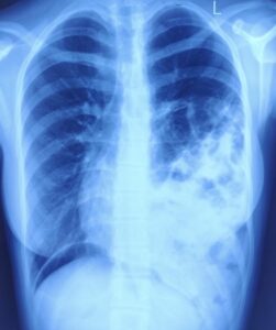
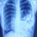
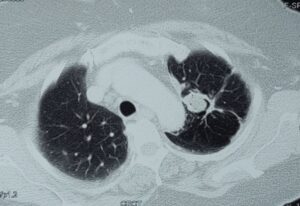
Surgery is performed using a High definition camera through small incisions which
reduces pain, morbidity leading to faster recovery.
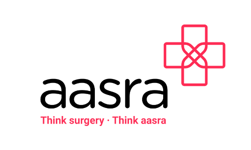
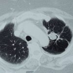
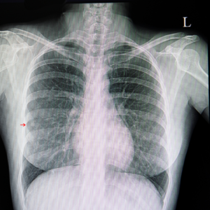
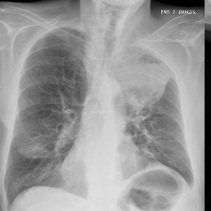
Surgery is performed by placing the incision between the ribs approximately 10-15 cm
long incision which can result in increased pain. Previously most of the procedures in the
chest cavity were performed using this method.
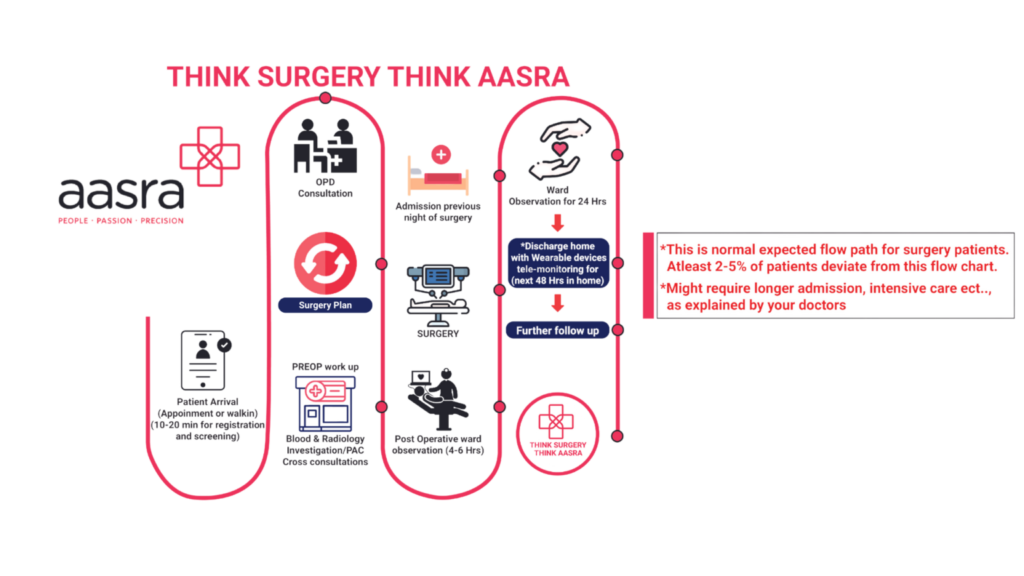
The recovery period is dependent on the complexity of surgery and also depends on the
type of disease. Usual recovery period is around 5-7 days on an average. Better to discuss with our Thoracic surgeon in person to get detailed plan of management.